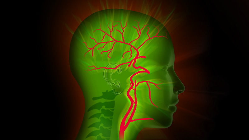

International studies have shown that CEA is the standard method for treating symptomatic carotid stenosis with over 70% congestion. During surgery, the plaque causing the stenosis is excised from the open artery. The artery is then closed with a patch. Blood flow is interrupted by temporarily clamping the carotid. However, care must be taken to avoid hypoxia in the affected hemisphere.
This can easily be monitored in regional anaesthesia. We all know the little yellow squeaky duck given to a cooperative and alert patient during vascular surgery. The patient squeezes the duck at regular intervals to let the surgeon know that brain activity is intact.

But what about patients under general anaesthesia?
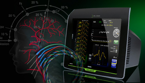
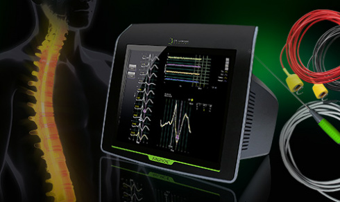
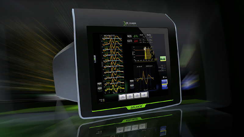
Somatosensory evoked potentials (SEPs) could be measured continuously by a neuromonitor.
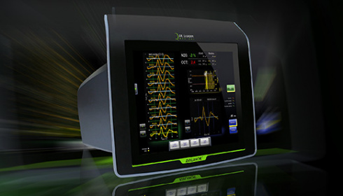
In order to use the AVALANCHE® SI neuromonitor in a CEA you need not be a trained neurophysiologist. Critical changes in the measured somatosensory evoked potentials will be picked up by our carotid programme and alert you in time of the risk of hypoxia. Over the years, equipment developed by Dr. Langer Medical have earned a reputation for ease of use and focusing on the essentials.
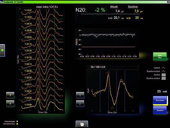
For the assessment of changes in the oxygen supply to the brain, somatosensory evoked potentials (SEPs) are measured continuously. Any changes are compared to the normal signal recorded just after anaesthesia was induced - the baseline signal.
If changes in amplitude and latency occur during the operation as a result of a reduced oxygen supply - something that happens in less than 16% of CEAs, an intraluminal shunt is needed.
This type of selective shunting reduces the post-operative stroke risk to below 17%. Intraoperative neuromonitoring does not pose any extra risk.
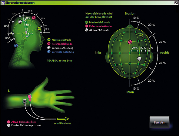
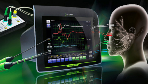
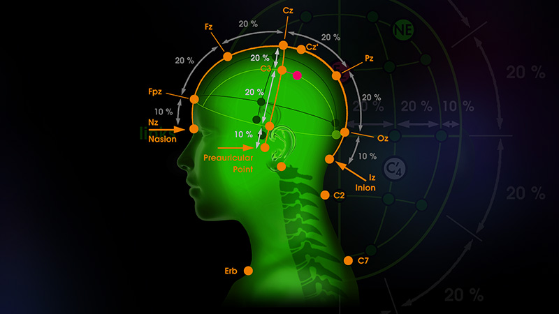
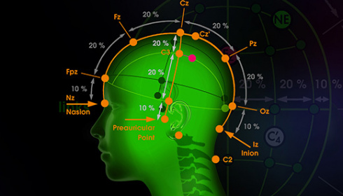
To stimulate the median nerve, surface electrodes are stuck to the wrist and connected to the stimulator.
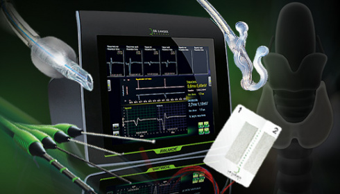
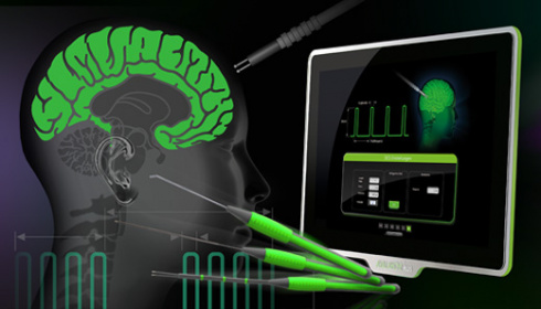
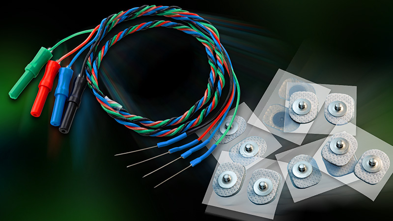
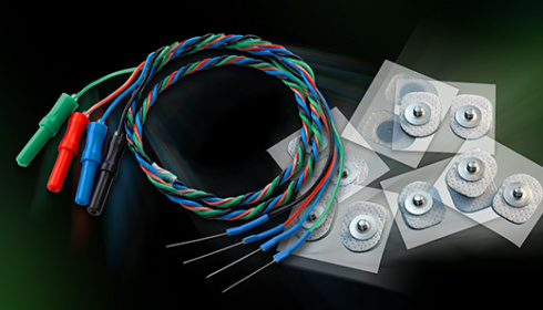
Whether one or several channels are required for the recording of EMG or EP signals or nerves and muscular tissue are to be stimulated, needle electrodes by Dr. Langer Medical GmbH will give you ample choice for neuromonitoring.
Various lengths, shapes and geometric arrangements as well as a palette of colours to suit your particular application purposes are available - the choice is yours.