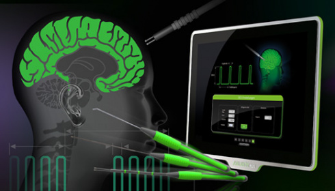

Intraoperative neuromonitoring of the cranial nerves and central nervous system is routine during neurosurgical interventions.
If a mass makes functionally important areas of the brain unidentifiable during surgery, AVALANCHE® PLUS can help you to find access to the tumour tissue with as little damage as possible, while preserving the brain areas. The function of many cranial nerves can also be controlled with the help of multimodal neuromonitoring
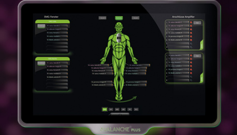
EMG - For testing a critical approach to motor nerves
Monitoring of motor cranial nerves (e.g. cranial nerves III-VII, IX-XII) by means of EMG is essential during surgery on the brain stem and skull base. An approach to motor structures can be detected promptly through continuous recording of muscle activity and acoustic reproduction of the free-running EMG. Stimulated EMG measurements facilitate functional control and localisation of the motor cranial nerves in the surgical site.
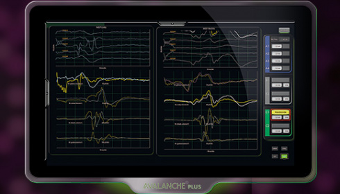
SEP - For monitoring sensory pathways and oxygen supply
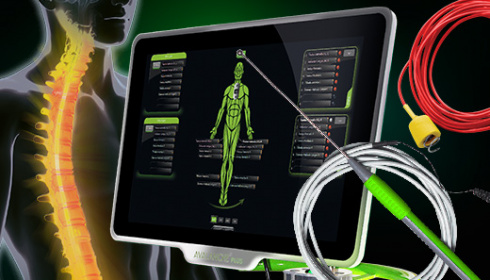
The continuous recording of somatosensory evoked potentials (SEP) provides important information about the functional state of the sensitive pathways. SEP measurement can, for example, serve to monitor a sufficient oxygen supply to the brain when clipping brain aneurysms – or the posterior funiculus of the spinal cord in the event of intramedullary tumours.
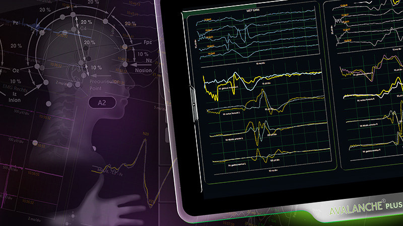
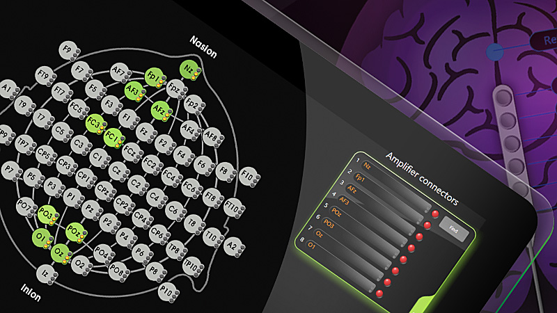
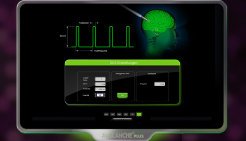
The AVALANCHE® PLUS SEP software offers predefined SEP leads. For example, you can select SEP stimulation on the median nerve at the touch of a button. No manual switching of recording sites required - AVALANCHE® PLUS automatically creates the measuring channel, displays the necessary recording sites in an anatomical view of the international 10-20 system and assigns a stimulation channel. All you need to do is assign the displayed recording site to a connection – quite simply by using the touch screen to drag and drop, while AVALANCHE® PLUS creates an SEP frame in the main window where you can monitor the SEP signals during surgery.
VEP - For monitoring the optic nerve
Intraoperative recording of visual evoked potentials (VEP) is used to monitor the function of the second cranial nerve (optic nerve) and includes monitoring the integrity of the visual pathway from the retina to the visual cortex. Reliable VEP recording can be a support in decision-making regarding the aggressiveness of tumour resection. Real-time adjustment of the strategic approach can reduce permanent, iatrogenic damage to the optic nerve. With AVALANCHE® PLUS VEP, the retina can be stimulated through the closed eye with LED flash goggles (red or white light). The sufficient intensity of the stimulation as a prerequisite for correct VEP measurement can be verified by simultaneously recording an electroretinogram (ERG).
MEP - For monitoring the function of central motor pathways
Measuring motor evoked potentials (MEP) triggered by transcranial or direct cortical stimulation can help monitor and protect motor pathways during almost all neurosurgical procedures by, for example, identifying motor structures or areas of the cortex, or functionally monitoring motor pathways (e.g. pyramidal tracts or motor cranial nerves).
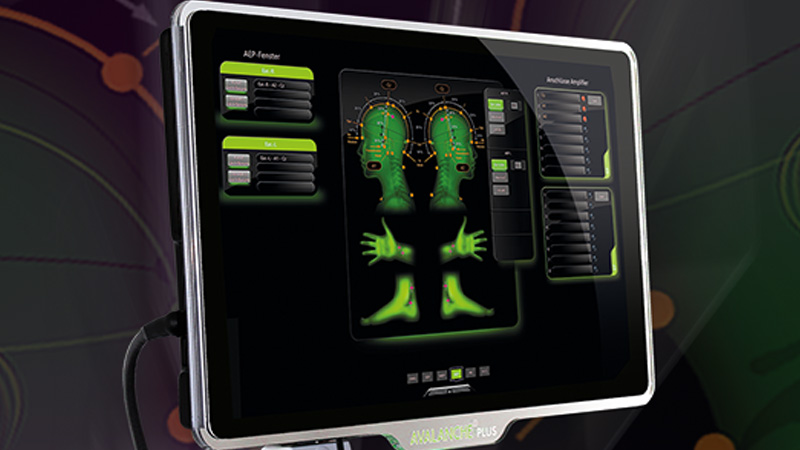
If you have already created an AVALANCHE® PLUS EMG configuration, there is hardly anything left to do for MEP, because the muscles have already been selected. You simply create an MEP measurement frame by assigning the muscles via drag and drop.
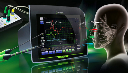
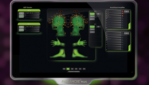
AEP - For monitoring the auditory nerve and brain stem
The auditory nerve as part of the vestibulochoclear nerve (cranial nerve VIII) is predominantly located in the cerebellopontine angle. Traumatic stress caused by manipulation around the cerebellopontine angle has a particular impact on this part of the auditory nerve. Maintaining and preserving auditory function is a significant aspect of intraoperative monitoring in cerebellopontine angle surgery, during which the patient is played pre-defined clicking stimuli through earphones.
The resulting action potentials are derived cortically. Automated monitoring of intraoperative changes in AEP potential can help to detect surgical manoeuvres that potentially compromise the patient’s auditory function. AEP potential monitoring provides the surgeon with direct feedback about the functional integrity of the auditory pathway.
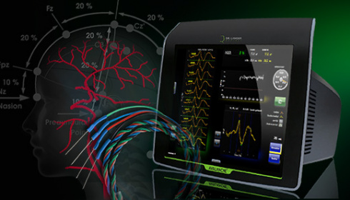
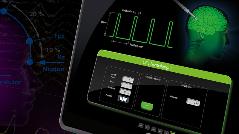
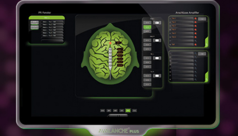
Direct cortical stimulation has been designed for localisation of motor areas in the brain and the speech centre in neurosurgery or epilepsy surgery (cortical mapping). Brain tumours can alter the anatomy of the cortex, which differs from patient to patient in any case, such that, without assistance, access to the tumour tissue with as little damage as possible to the surrounding tissue while preserving functionally vital areas of the brain is difficult. With AVALANCHE® PLUS, it is possible to visualise a “map” of the patient's individual cortex and localise the regions of interest. Special atraumatic stimulation probes are available for stimulation.
Whether Penfield or train technique, AVALANCHE® PLUS graphically illustrates complex stimulation parameters for direct cortical stimulation. No matter which parameter you change, it is all clarified immediately. Full functionality paired with simplest handling.
AVALANCHE® PLUS makes you a specialist!
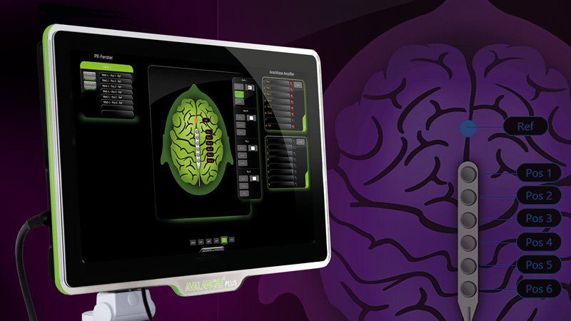
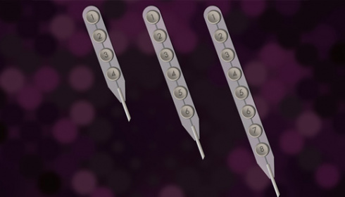
Tumours in the central cortex can alter the anatomy to such an extent that it is imperative to localise the structures in the area of the central sulcus in order to decide on the type and extent of tumour resection. SEP phase reversal measurements can be used to identify the central cerebral sulcus. The signals usually generated by stimulation at the wrist (median nerve) are measured directly at the cortex with strip electrodes. A polarity change in the evoked potential across the central sulcus can be deduced from the SEP curves, which corresponds to the “phase reversal” between motor and sensory cortex. This can spare healthy tissue and reduce deficits.
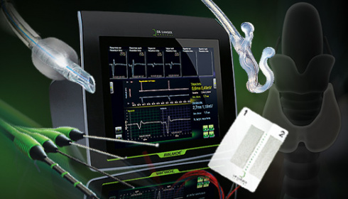
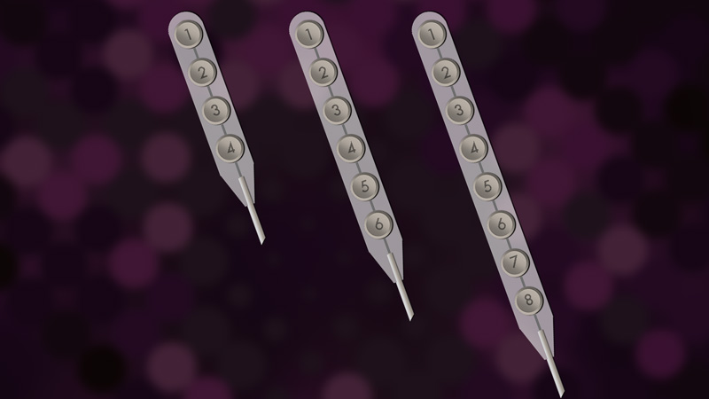
Select strip electrodes with 4, 6 or 8 contacts with a single click in SEP phase reversal and let AVALANCHE® PLUS automatically take care of the rest: Electrode visualisation, phase reversal window and phase reversal calculation – complex measurements configured in an instant!
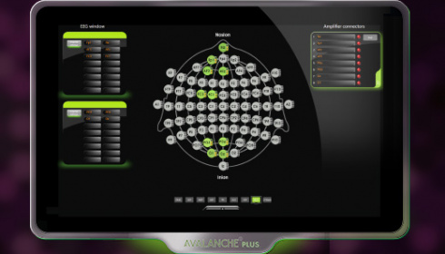
Electrical brain activity can be measured intraoperatively via the scalp (EEG - electroencephalography) or direct cortically (ECoG - electrocorticography) via subdural strip electrodes. These real-time insights into spontaneous brain activity help the surgeon, for example, to delineate epileptic foci or to detect seizures during direct cortical stimulation. In combination with other IONM modalities, EEG recording can be used to monitor cerebral perfusion.
The AVALANCHE® PLUS system utilizes EEG for precise monitoring during various neurosurgical procedures using needle electrodes or strip electrodes (ECoG) to provide real-time assessment of brain activity and make surgical interventions effective.
The AVALANCHE® PLUS makes you a specialist!
Detection of the central sulcus with AVALANCHE® PLUS
Phase reversal SEP measurements can be used to identify the central sulcus. The potentials evoked by stimulation at the wrist (median nerve) are detected directy at the cortex using strip electrodes.
Contact spacing 10 mm, cable length 1.85 m, with connectors according to DIN 42802-1, single sterile packed, disposable, PE = 1 pc.