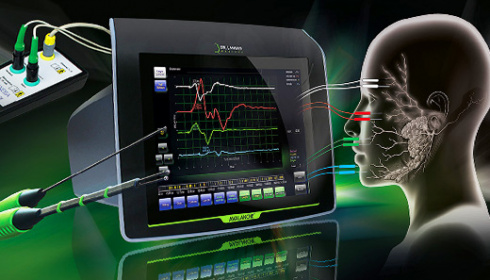


Although tumours of the parotid gland are less common than other tumours, several thousands of operations are carried out in Germany every year to partially or entirely remove the parotid gland. The risk of damaging the delicate branches of the facialis nerve is substantial. Damage to just one of those delicate structures during surgery may mean a disastrous outcome for the patient. Immediate physical consequences such as failure of closing an eyelid or a drooping corner of the mouth, alterations in the sense of taste, or reduced tear and saliva secretion often affect the psychological well-being of the patient.
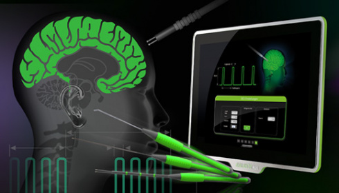
Intraoperative neuromonitoring (IONM) using AVALANCHE® SI helps to identify motoneurons and monitor their function during surgery.
Fields of application
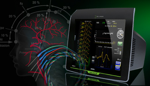
Identification and function check of the facialis nerve in parotid surgery, mastoidectomy, insertion of cochlea implants and middle ear surgery.
Monitoring of cranial motor nerves during tumour resection in the oral cavity, ear, neck, nose and the base of the skull.
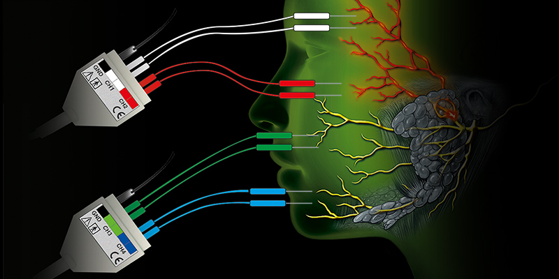
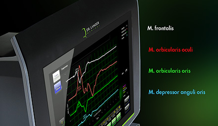
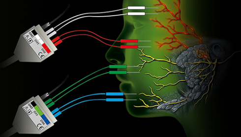
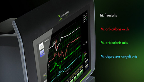
During intraoperative neuromonitoring, minute electric impulses are transmitted to the nerves by special stimulation probes. The intact nerves conduct the excitation to the corresponding facial muscles, where the generated muscle action potential is measured through electromyography (EMG) and converted into visual or acoustic signals.
Four EMG signal graphs in distinct colours show the functioning of the main branches of the facialis nerve. They will show up the slightest irritation of a nerve. Unintentional pressure or traction, stretching or compressing a nerve, will result in the spontaneous contraction of the muscle affected. AVALANCHE® SI picks up the impulse and sends out acoustic or visual hints.
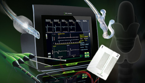
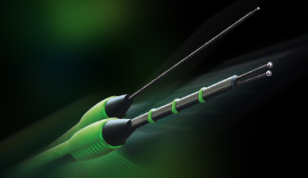
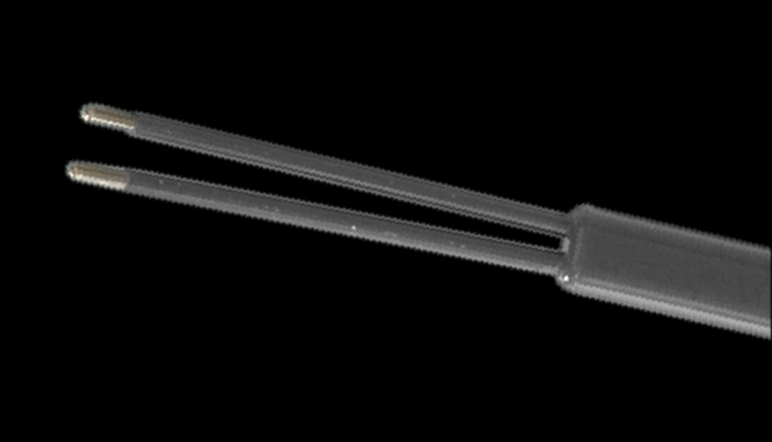
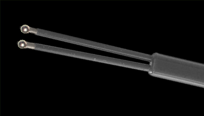
With some types of surgery, a simple option of monitoring neural function is sufficient. A distinct twitch of the target muscle upon nerve stimulation is all that is needed to know that everything is fine. An assistant may be given the task of observing the response of the target muscle to electrical stimulation. Thus, the surgeon can focus entirely on the operation site without losing sight of the sensory structures. In this context, it can be helpful to connect two stimulation probes simultaneously, for example a bipolar and a monopolar stimulation probe.
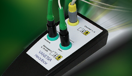
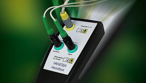
The headbox can be hooked onto the operating table. It has two stimulation ports so that you can use monopolar or bipolar stimulation without having to change probes.
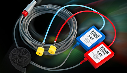
Extend your AVALANCHE® SI by adding a measuring system for auditory evoked potentials (AEP) for surgery where the auditory function is all-important. EMG and AEP combined in a single portable neuromonitor - this is absolutely unique!
The choice is yours: Bipolar or monopolar, conventional or minimally invasive, with or without microscope.
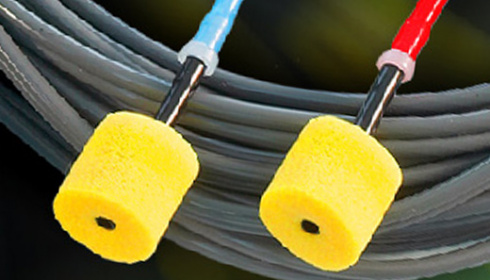
Reusable or disposable accessories -
Our range offers a matching solution for virtually all situations.
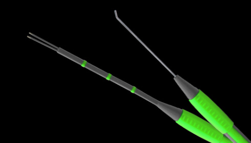
Our stimulation probes have been made with special attention to ergonomics.
Quality made in Germany by Dr. Langer Medical.
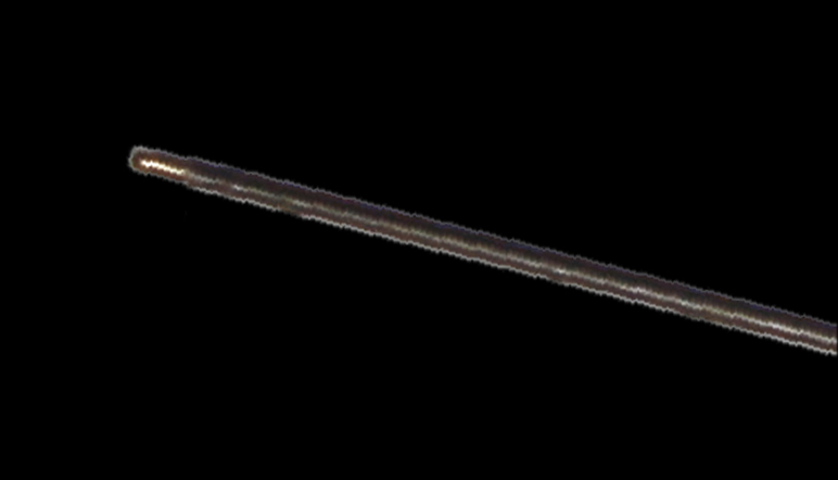
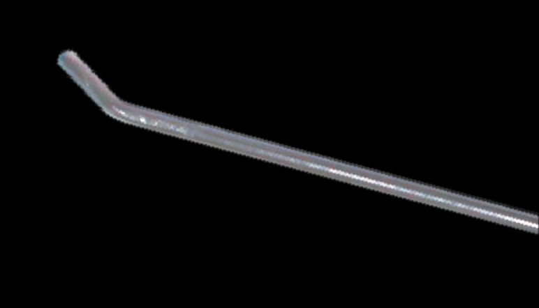
Whether one or several channels are required for the recording of EMG or EP signals or nerves and muscular tissue are to be stimulated, needle electrodes by Dr. Langer Medical GmbH will give you ample choice for neuromonitoring.
Various lengths, shapes and geometric arrangements as well as a palette of colours to suit your particular application purposes are available - the choice is yours.