the art of neuromonitoring
in spine surgerySituation
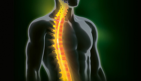
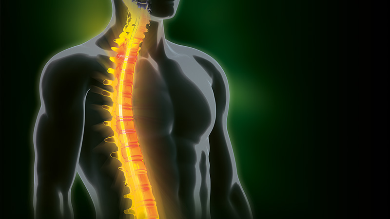
Neuromonitoring in spine surgeryThe spinal cord includes the descending motor nerves that mainly carry impulses from the brain to the muscles, as well as the ascending sensory pathways that transmit information from the peripheral sensory organs to the brain.
There is a general distinction between neuromonitoring of efferent (descending) motor pathways via EMG (electromyography) and motor evoked potentials (MEP), and neuromonitoring of afferent (ascending) pathways using somatosensory evoked potentials (SEP).
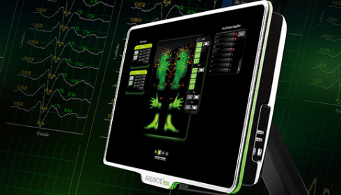
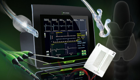
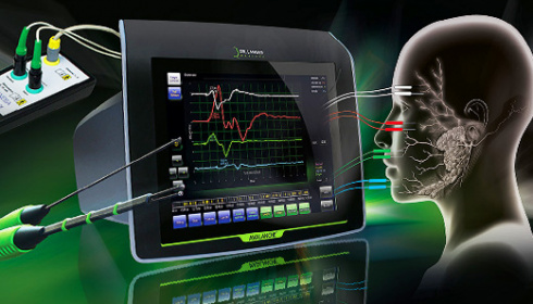
What at first glance seems very complex, could be extremely easy with our AVALANCHE® PLUS. In surgical scenarios, where a combination of the different methods is required and measurements are taken simultaneously, AVALANCHE® PLUS has a completely new approach.
Application
Spinal surgery e.g. in scoliosis, kyphosis, decompressions, degenerative spinal diseases, discectomy, during the resection of spinal tumours or for inserting pedicle screws etc.
AVALANCHE® PLUS EMG
for the monitoring of critical proximity to motor pathwaysWhen measuring electromyograms (EMG), motor nerves in the operation site are electrically stimulated. The evoked action potentials are transmitted to the peripheral muscles, then picked up and measured by electrodes and visualized on the neuromonitor.
Continuous EMG recording in several muscles simultaneously enables surgeons to respond immediately when they come critically close to motor neural pathways and signal activity increases.
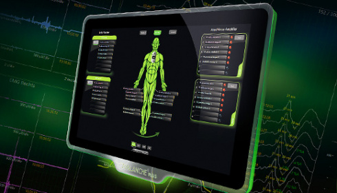
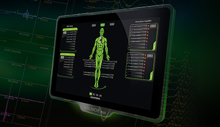
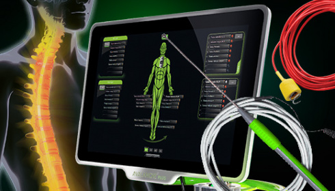
The selection of muscle groups and the required number of channels depends on the surgical scenario. Time consuming entering of muscles via the keyboard is no longer necessary. Setup in AVALANCHE® PLUS is easily done with drag and drop via the touch screen.
AVALANCHE® PLUS comes with predefined muscle groups for every spinal cord level in an anatomic overview. Simply select individual muscle groups and assign them to the desired measuring window. Once you assign the connector, EMG configuration is complete.
Pedicle Screw Testing
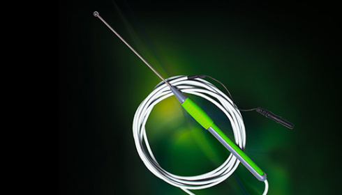
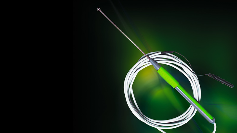
with AVALANCHE® PLUS and the monopolar ball probeSpecific monopolar stimulation probes are available for the insertion of pedicle screws. These permit stimulation within the bore hole itself or through surgical instruments during the drilling and the screwing process. If the stimulation results in an increased signal activity on the neuromonitor, caution is advised – there could be nerve compression or perforation of the pedicle wall.
In the AVALANCHE® PLUS software activation of the pedicle screw stimulator is done by a push of a button. The user interface is identically designed as the AVALANCHE® PLUS I/O-Box. The probe connector on the AVALANCHE® PLUS I/O-Box is colour and shape coded – confusion is nearly impossible.
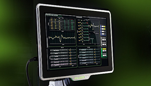
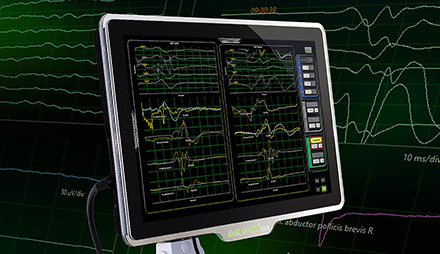
In the main window you are able to display either all measuring windows of all modalities or one modality only. For monitoring of pedicle screws, choose the EMG view and keep all essential signal changes in view on the large touch screen.
AVALANCHE® PLUS MEP
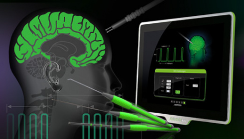
for the functional check of central motor pathwaysMotor evoked potentials (MEPs) are a particular form of evoked potentials, measured through an EMG. They are triggered by transcranial or direct cortical stimulation, and the response signals are picked up at the corresponding extremity muscles. This enables the surgeon to quickly assess the state and functionality of the spinal cord.
If you have already implemented an AVALANCHE® PLUS EMG setup, MEP configuration is nearly finished. The selection of muscle groups is already done. Just create a MEP measuring window by assigning the muscle groups via drag and drop – setup can be that easy.
If stimulation pulses are released by the transcranial stimulator, AVALANCHE® PLUS triggers the MEP measurement automatically. MEP signals are display in the measuring window without any need of user action. Choose the MEP view to monitor critical phases in surgery comfortable.
AVALANCHE® PLUS SEP
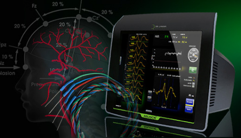
for the monitoring of ascending pathways of the spinal cordTo ensure that changes that could result in post-operative neurological deficits, somatosensory evoked potentials (SEPs) are continually measured. Signals picked up in real time are compared to the normal signal recorded just after anaesthesia was induced - the baseline signal.
The signal generated by electrical stimulation of the median nerve at the wrist or at the tibial nerve at the foot is acquired as an SEP cortically or subcortically and shows typical changes in amplitude and latency. These alert to potential or actual threats for the nerve during operation.
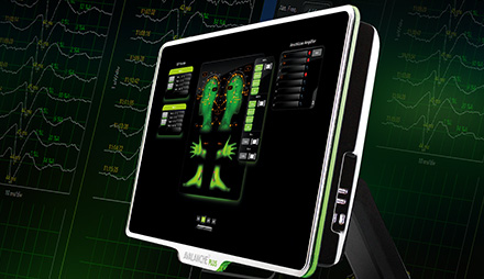
The AVALANCHE® PLUS SEP software comes with predefined SEP channels. At the push of a button, SEP stimulation of the median nerve is chosen. Creating the signal channel manually does no longer exist - AVALANCHE® PLUS creates the signal channel automatically, highlights the required positions of the international 10-20-system in an anatomic overview and selects the stimulation channel by itself.
Assign an electrode connector to the highlighted position of the 10-20 system – easily via drag and drop on the touch screen. In the meantime AVALANCHE® PLUS generated a SEP measuring window to monitor all SEP signals during surgery.
Stimulation probes
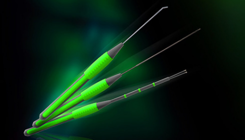
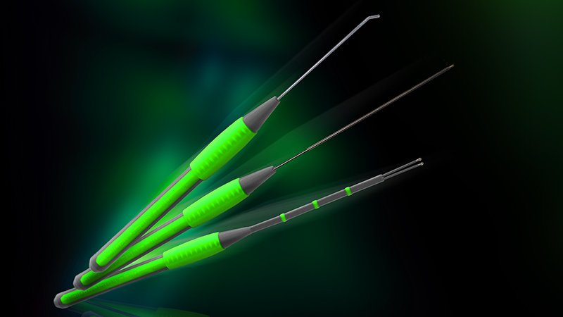
Electric stimulation of nerves and neuronal structuresThe choice is yours: Bipolar or monopolar, conventional or minimally invasive, with or without microscope. Reusable or disposable accessories -
Our range offers a matching solution for virtually all situations.
Our stimulation probes have been made with special attention to ergonomics.
Quality made in Germany by Dr. Langer Medical.
Needle electrodes
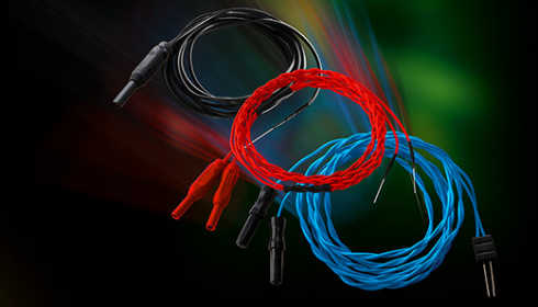
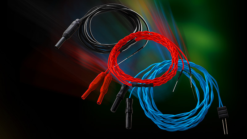
Colour-coded needle electrodes for recording and stimulationWhether one or several channels are required for the recording of EMG or EP signals or nerves and muscular tissue are to be stimulated, needle electrodes by Dr. Langer Medical GmbH will give you ample choice for neuromonitoring.
Various lengths, shapes and geometric arrangements as well as a palette of colours to suit your particular application purposes are available - the choice is yours.

